
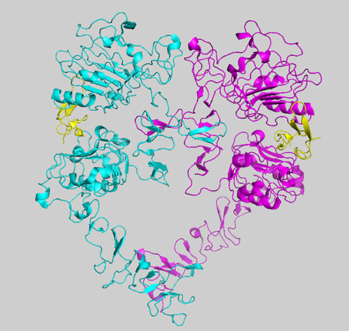
The EGF receptor is a classical receptor tyrosine kinase composed of an extracellular ligand binding domain, a single pass alpha helical transmembrane segment, an intracellular kinase domain and an unstructured C-terminal tail. In the absence of ligand, the EGF receptor exists as a monomer. However, upon binding EGF, the receptor dimerizes. This leads to the formation of an asymmetric dimer of the intracellular kinase domains, which results in the activation of the kinase. The receptor phosphorylates tyrosine residues on its C-terminal tail. These phosphotyrosines then serve as binding sites for the SH2- and PTB domain-containing signaling proteins that mediate the intracellular effects of EGF.
The EGF receptor is the first member of the ErbB family of homologous receptors. Besides the EGF receptor, the ErbB family includes ErbB2, ErbB3 and ErbB4. All the ErbB receptors can interact with other family members. Thus, there are at least eight different homo- or heterodimers that can form. The properties of these dimers differ as the characteristics of the various family members are different. For example, ErbB2 has no known ligand and thus is activated solely by forming heterodimers with one of the three ligand-binding members of the ErbB family. ErbB3 binds a ligand but its kinase is inactive. Thus, it is obligated to heterodimerize with a kinase-active family member, typically ErbB2, to generate a signal.
Mutation or over-expression of the ErbB family members is associated with several different types of cancer, including breast cancer, lung cancer and glioblastoma. Thus, understanding how these receptors function within the context of the overall signaling network is key to developing effective therapies for many different types of cancer.
The goal of my research is to identify structure-function relationships within the ErbB family of receptors to understand what drives their interaction and how this is regulated. We are also interested in how the receptors mediate the earliest steps in the process of signal transduction. The work in my laboratory revolves around several issues.
I. Signaling via the C-terminal tail. The C-terminal tail of the EGF receptor (and all ErbB receptors) is intrinsically disordered, a property shared by many other proteins that are hubs in a signaling network. Within the C-terminal tail there are nine phosphorylatable tyrosine residues that lie in linear motifs that are recognized by dozens of different SH2- or PTB domain-containing proteins. Questions we are addressing are: How many signaling proteins can be bound by a single EGF receptor at once? How does the binding of one signaling protein affect the ability of the tail to bind a second signaling protein? How does the intrinsically disordered structure of the tail relate to its ability to bind so many different signaling proteins. Is the ability of the C-terminal tail to bind proteins dependent only on the linear phosphotyrosine motifs or are there other auxiliary sequences that contribute to protein binding. We are using mutational analyses, MD simulations and mass spec footprinting to answer these questions.
II. Agonist-dependent differences in signaling. The EGF receptor has seven different ligands. Studies have shown that different ligands can elicit different effects in a single cell even though they bind to the same receptor. The question is how is this accomplished. To address this, we are using luciferase fragment complementation imaging combined with principal component analysis to characterize and compare the signaling networks utilized by the different EGF receptor agonists. We are also using radioligand binding studies to identify differences in the way different ligands interact with the EGF receptor.
III. The ErbB2/ErbB3 heterodimer. The ErbB2/ErbB3 heterodimer is the most highly transforming ErbB dimer. Yet little is known about how the dimer is assembled, whether tetramerization of the receptors occurs and how the heterodimer signals given that one partner, ErbB3 is kinase-impaired. We are using mutational analyses and radioligand binding studies to characterize the interaction between ErbB2 and ErbB3. We will also be using luciferase fragment complementation imaging to determine how the binding of signaling molecules to the ErbB2/ErbB3 heterodimer differs from their binding to the EGF receptor homodimer.
Research | People | Publications | Methods | Teaching | Links | Home