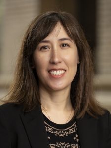
Spotlight on Faculty – Robertson, Janice
 “Life began with little bags of garbage,” proposed the physicist Freeman Dyson. “Membranes made of oily scum […] enclosing volumes of dirty water containing miscellaneous garbage.” Billions of years of evolution have shaped cell membranes from simple “bags” into complex and finely-tunable structures. The cell membrane is not just a barrier separating the chaotic extracellular environment from the controlled intracellular space; they allow for the storage of electrical and chemical potential energy, facilitate transport of substances into and out of the cell, and change the cell’s shape according to its biological needs.
Key to these functions are the proteins embedded within the cell membrane. While much is known about how proteins self-assemble in water—changing from a string of polypeptides into a folded, functional shape—relatively little is known about how this is done in the cell membrane. Dr. Janice Robertson, Assistant Professor of Biochemistry and Molecular Biophysics, and her lab use single-molecule techniques and computational modeling to find answers to the questions surrounding membrane protein assembly.
“Protein sequences have non-polar, or ‘greasy,’ residues scattered amongst the sequence,” said Robertson. “These greasy residues want to form a little oil droplet to shield themselves away from water.” When these residues come together, they support the folding of the sequence into the protein’s defined shaped. But the cell membrane, a lipid bilayer, is a completely different environment from water. It’s essentially a very thin, defined layer of oil. “So then the question is why do these residues come together into a folded structure? How do greasy protein surfaces choose other greasy protein surfaces instead of the similarly greasy membrane that surrounds it? How do you hide oil from oil? This is what we’re trying to figure out.”
Today, the concept of membrane proteins is taken for granted, but the idea wasn’t widely accepted until the 1970s. “They thought, no you can’t put proteins in the membrane, that would just dissolve it,” said Robertson. “It’s kind of remarkable that we didn’t really think that membrane proteins existed before my lifetime.”
Despite accounting for about 25% of proteins, membrane protein research remains decades behind water soluble proteins. This is largely because membranes are much more difficult to work with than water. “The big problems are knowing if you change the system in some way—like changing the lipids in the bilayer—if that’s doing anything else to your system,” said Robertson. “Lipids can segregate into patches of one type of lipid versus another and that is going to change the free energy of your system. Water is isotropic; you’re not going to have little pockets of ice forming at room temperature. But you can have that in a membrane. You have to be aware of that, and be very humble to the membrane.”
To get at these questions of how membrane protein structures come together, the Robertson Lab uses CLC, a family of chloride channels, as a model system. CLCs are large protein complexes made up of two subunits, and each subunit has one pore. This forms a so-called “double-barreled shotgun” for transporting chloride across the membrane. Whenever the two subunits are observed naturally, they’re seen together as a dimer. However, when diluted they can be seen as dissociated monomers which retain their function. This independence means that the dimerization is not a fixed structure required for proper function, but a dynamic process—one that can be manipulated with different lipid compositions. By studying the conditions that bring CLC subunits together, and those that keep them apart, Robertson and her lab can gain insights into how other large protein structures come together in the membrane.
However, imaging these systems at such low dilutions cannot be done in the conventional way. They require single-molecule approaches. “So we built our own single-molecule fluorescence microscope for imaging membrane proteins,” said Robertson. The custom microscope is an improvement over the TIRF (Total Internal Reflection Fluorescence) design, making it a versatile, multi-colored co-localization system. This allows Robertson and her lab the ability to see the single-molecule reaction occurring right in front of their eyes. “We have the potential to see dynamic binding and unbinding, or we can see this whole thing moving together in the membrane.”
Understanding these systems requires both complex computational and experimental studies—which Robertson is uniquely qualified to lead. “For my Ph.D., I took what people think of as a non-conventional pathway,” said Robertson. “I did basically entirely computer modeling. And then I knew I didn’t want to be limited in my computational studies by the experiments that were available. I wanted to be doing the experiments myself. So then I did a post-doc in one of the best experimental membrane protein biochemistry labs. I went from doing 100% computer studies to 100% experiments—but I didn’t have any experience. It was like I was an undergraduate student again, immersed in a completely new environment. But I had the ability to just say, ‘I don’t know this system; please teach me and I will learn everything.'”
Following 5 years at the University of Iowa—fine-tuning the experimental techniques and building the microscope—Robertson moved her lab here, to the Department of Biochemistry and Molecular Biophysics at Washington University in St. Louis, in September 2018. With the experimental technique established, the Robertson Lab is poised to combine it synergistically with computational modeling. “And for the next five years, we’re going to be working in both directions,” said Robertson. “The hope is that we’re going to get a rigorous and comprehensive understanding of what is going on in the membrane.”
There are still many questions surrounding the physics of membrane protein folding, but recent advances have injected the field with excitement and growth. “I hope that at the end of my career we’ll have some of the answers,” said Robertson. “Certainly, we’ll have many more questions, but I hope that we can map out the fundamentals so there’s a solid field from which a lot of people can do a lot of interesting research.”
Written by Kevin Blake, 22 Aug 2019
Kevin Blake is a second-year graduate student, in the lab of Dr. Gautam Dantas, and the Plant and Microbial Biosciences (PMB) Ph.D. program.
