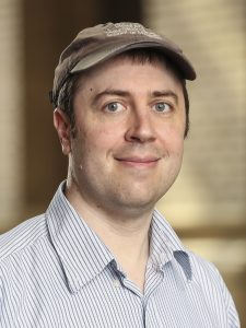
Spotlight on Faculty – Greenberg, Michael
The Heart of Research
As we uncover previously unknown truths about the world surrounding us, scientific research becomes more complex making interdisciplinary research vital to the progress of human health.
Dr. Michael Greenberg is an Assistant Professor of Biochemistry and Molecular Biophysics who has made the very core of his lab interdisciplinary in nature. Greenberg’s lab is at the epicenter of biochemistry, biophysics, and cellular biology. His lab studies cytoskeletal motors using physics to explain cellular movements.
His interest in the intersection of these fields started as early as his undergraduate experience at Brandeis University where he triple-majored in physics, chemistry, and biological physics.
“Initially I thought I was going to study string theory,” Greenberg said. “I realized pretty quickly that I didn’t want to sit with a pencil and paper all day. For me, it was more fun to learn about string theory than it was to do it on your own.”
From there, Greenberg spent his graduate career and postdoctoral fellowship studying molecular motor mechanics with Dr. Jeffrey Moore and Dr. Michael Ostap, respectively. These experiences collectively inspired him to begin his own lab studying how cytoskeletal motors function in health and disease. His goal is to explain the way cells and molecules move using physics and modeling. However, before starting his own lab, Greenberg had little experience with cell culture.
“There are lots of really interesting questions in biology for people with physics backgrounds to tackle,” Greenberg said. “We use physics-based models and techniques to try to understand biological systems.”
In his pursuit to understand how cells and tissues move at a molecular level, Greenberg’s lab has become adept in utilizing human engineered heart tissues. These model tissues are three-dimensional, and have the ability to contract similar to a human heart. Greenberg’s lab builds the human engineered heart tissues from scratch using Matrigel, collagen, stem cell-derived cardiomyocytes, and stem cell-derived fibroblasts.
“The tissues will self-assemble between two sticks and they will contract,” Greenberg said. “We can take two electrodes, put them on the sides of the tissue, and hook them up to a signal generator. We can also take these tissues and apply mechanical loads to see how that affects contractility.”
When reports of SARS-CoV-2 patients dying of heart failure reached the department, Greenberg, Dr. Kory Lavine (Department of Medicine; Division of Cardiology), and Dr. Michael Diamond (Department of Medicine; Division of Infectious Diseases) began collaborating to find out why.
However, the team faced many challenges working with SARS-CoV-2 in a biosafety level three facility. In such a high biosafety level facility, there are strict rules about what can and cannot be taken in and out of the laboratory. The team had to get creative with ways to measure contractility of the human engineered heart tissues without a recording video microscope. Using a simple camera, a monitor on a conventional microscope, a selfie stick, and some novel software written by Greenberg himself, the team showed that stem cell-derived cardiomyocytes can be infected with SARS-CoV-2 which causes them to decrease contractility similar to what is seen in failing heart tissue.
“It was a great project to get involved with, and a fantastic collaboration because we had three groups with three very different areas of expertise,” Greenberg said. “I think we were able to come together and do something really cool.”
Their work, entitled SARS-CoV-2 Infects Human Engineered Heart Tissues and Models COVID-19 Myocarditis, is currently available as a preprint on bioRxiv. Greenberg looks forward to continuing to do research on the cusp of cellular biology and physics to explain human health.
“Cells are at the point where we can manipulate them and do a lot of things that [in the past] we could only study biochemically in vitro,” Greenberg said. “We can test mechanistic hypotheses about these complex systems from single molecules to higher order levels of organization.”
Outside of the lab, Greenberg enjoys spending time outdoors with his four children, learning about the forces behind the weather such as tornadoes and hurricanes, and playing heavy metal music on his guitar. In addition, he loves to cook.
“There are a lot of similarities between cooking and doing wet lab; however, cooking has a much higher frequency of success,” Greenberg said.
 For over 70 years, the anticoagulant Warfarin has been a first line defense used to treat and prevent heart attacks, strokes, and other cardiovascular diseases. Warfarin inhibits the Human Vitamin K Epoxide Reductase (VKOR) to hinder coagulation. However, despite being the most popular prescribed anticoagulant, Warfarin has a very narrow range of efficacy and overdose often induces severe, sometimes fatal bleeding. In older adults, one-third of hospitalization for adverse drug reaction are due to warfarin use.
 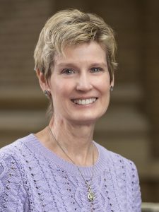 Snapshot of a Scientist
 Illuminating the Invisible: The Soranno Lab’s Study of APOE4 Conformations
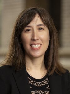 “Life began with little bags of garbage,” proposed the physicist Freeman Dyson. “Membranes made of oily scum […] enclosing volumes of dirty water containing miscellaneous garbage.” Billions of years of evolution have shaped cell membranes from simple “bags” into complex and finely-tunable structures. The cell membrane is not just a barrier separating the chaotic extracellular environment from the controlled intracellular space; they allow for the storage of electrical and chemical potential energy, facilitate transport of substances into and out of the cell, and change the cell’s shape according to its biological needs.
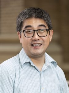
 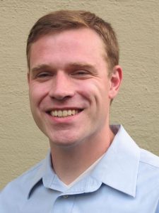 When Greg Bowman presents a slideshow about the proteins he studies, their 3D shapes and folding patterns play out as animations on a big screen. As he describes these molecules, it might be easy to miss the fact that he can’t really see his own presentation, at least not the way the audience does.
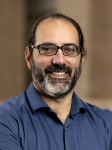
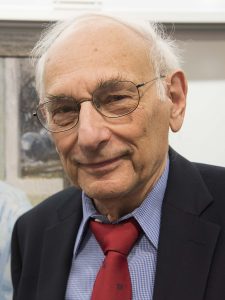 Carl Frieden was born and raised in New Rochelle, New York. He obtained his B.A. degree from Carleton College and his Ph.D. in Chemistry from the University of Wisconsin under the mentorship of Robert A. Alberty. In 1955, Dr. Frieden joined the Department of Biological Chemistry at Washington University School of Medicine as a postdoctoral fellow to work with Dr. Sidney Velick. He was hired by Carl Cori as an Instructor in 1957 and promoted to full professor in 1967. Dr. Frieden has served both as Interim Head (from 1987 to 1990 and from 1997 to 2000) and Head (from 2000 to 2005) of the Department. Among other awards, he has received the Carl and Gerty Cori Faculty Achievement Award and the Second Century Award from Washington University. He has also been awarded the Peter Raven Lifetime Achievement Award from the St. Louis Science Center as well as the Christian B. Anfinsen Award from the Protein Society. He was elected to the National Academy of Sciences in 1988 and to the American Academy of Arts and Sciences in 2004. From 1995-2000 he was an Alumni Endowed Professor and, from 2000 to 2005, the Wittcoff Endowed Chair of the Department of Biochemistry and Molecular Biophysics. He served as Director of the Medical Science Training Program (MSTP) from 1986-1991.
 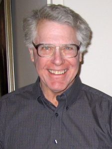 Stepping into Elliot Elson’s lab is like falling into the rabbit hole in Alice in Wonderland. There are movable amplifiers perched precariously on a sled of steel runners, lasers in black boxes, pieces of salvaged equipment, cords and cables spewing like spaghetti from another machine, and a huge black tent that looks like it was inspired by Darth Vader. This is a workshop for scientific invention. The tunnel burrows deeper into the natural world’s mysteries. The elusive White Rabbit entices us to follow and to probe the enigmas of life. Elliot Elson seeks to understand and characterize cells and tissues as physical entities. His “white rabbit” has led him to adventures and discoveries that have yielded new insights into the inner workings of life itself—in several different key fields.